A baby’s head shape is not always perfectly round. Here, a pediatrican walks us through infant head shape development and possible medical concerns from newborn to toddler.
The information in this article is provided by Pediatrician Leah Alexander. The purpose is to provide examples and knowledge to help parents dig deeper into their baby’s situation, not to offer a complete picture or a possible diagnosis. Medical concerns should always be handled by a doctor who meets the patient.
Infants do not always have perfectly round-shaped heads, and it is easy to start worrying as a new parent.
Why does my baby have such an odd head shape? Will they ever look normal? Is the uneven shape a sign of some underlying condition?
The head of a newborn differs from that of an older infant or toddler. Premature infants have softer skull bones than full-term infants. In addition, some factors can change a baby’s head shape. Some head shapes may be a sign of a problem or underlying medical condition. It is important to distinguish what shape is normal from what is a sign of a problem.
To sort out what’s normal and not, we asked Pediatrician Leah Alexander to walk us through infant head shape development and possible medical concerns.
Your Baby’s Head Shape: Normal Development, Medical Concerns
The Anatomy of an Infant’s Head
An infant’s head consists of an outer layer of hairy skin, underlying muscle, and skull bones. Inside the skull lies the brain with its connecting nerves and blood vessels. The brain is surrounded by a liquid called the cerebral spinal fluid. This fluid both cushions the brain and provides a conduit for nutrients and the removal of cellular waste.
At birth, the skull is separated into five bone pieces that are held together by connective tissue. This structure is important to allow mobility of the skull bones as the baby’s head passes through the birth canal. It also leaves room for the brain to grow over the first few years of life. The open spaces between the bones are called fontanelles. Most newborns have four of these “soft spots”, the largest on the top of the head.
Over time, the skull bones grow, harden, and come together to form a complete vessel for the brain.
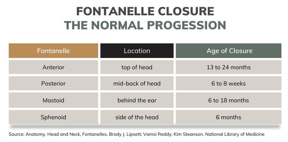
The Normal Progression of Fontanelle Closure
Each fontanelle closes at a different time throughout the first two years of life. The size of the anterior and posterior fontanelles are typically larger among infants of African ancestry. The anterior fontanelle closes earlier in boys than it does in girls.
If any of these openings close too soon, the skull can restrict brain growth. Any that remain open longer than expected can indicate a head size that is too large.
At routine well-visits, the doctor will examine your baby’s head to check for signs of a problem. Its size will be measured and recorded on a head circumference chart. This allows any problems to be detected early. What is considered a “normal” head size differs for each infant and is influenced by hereditary factors.
It is more important for the head to grow at a steady and consistent rate and for it to be proportional to the size of the infant’s body.
Reasons For Abnormal Infant Head Shapes
Birth-related Causes
Prolonged Delivery
Sometimes, the birth process itself alters the shape of a newborn’s head. When a vaginal delivery takes longer than expected, the infant’s head can become molded within the birth canal. This may result in what some parents call a “cone-shaped head.”
Fortunately, this is a temporary issue. The soft connective tissues and flexible skull bones return to a normal shape during the first week of life.
Vacuum Extraction
If an assistive device is used during the delivery to help the infant’s head pass, the pressure can result in a temporary head-shape abnormality. In most cases, a vacuum device is used, attaching it to the infant’s head to pull as the mother pushes. It is used when fetal distress occurs and the baby must be delivered quickly. It may also prevent a cesarean section.
After a vacuum procedure, the baby may have a “cone-shaped” head due to swelling under the scalp from the suction force of the vacuum device. This swelling, called a caput succedaneum, self-resolves within the first two days of life.
Alternatively, an accumulation of blood may form under the scalp, called a cephalohematoma. A difficult delivery of an infant with a large head may cause it as well. Unlike a caput succedaneum, this collection of blood is typically located on one side of the head. It can take one to two months for a cephalohematoma to go away. As the blood breaks down, it may cause the baby’s skin to appear yellow or jaundiced.
While still in the hospital, doctors may order a blood test to make sure these blood byproducts are not at a high level. The size of the cephalohematoma will gradually decrease as the blood within it is reabsorbed and metabolized. Larger ones, however, may take more time to resolve and can calcify, creating a permanent change in the shape of the skull.
Head Shape Abnormalities Associated with Positioning
Plagiocephaly
During the first few months of life, parents may notice that an area of their baby’s head begins to appear flat. This head shape change is known as plagiocephaly. It most often occurs as the baby’s head rests on a flat surface for extended periods of time.
The back of the head is the most common location because of the amount of time infants spend lying on their backs. This type is more specifically called brachycephaly. By age three months, 46 percent of infants have some amount of head flattening. It may, however, be present at birth in newborn twins or multiples due to in-utero pressure against the head.
Some infants develop flattening one side of the head along with facial asymmetry. These infants tend to look more in one direction than the other, resulting in pressure on that same side of the head. Usually, the baby can easily turn the head in either direction, but there is a preference for one side.
Some infants, however, have a neck muscle problem that limits head movements. If a right or left sternocleidomastoid muscle is injured during birth, it can result in torticollis. In such situations, intervention is necessary to improve neck mobility and prevent longterm problems.
Because of weeks to months of neonatal intensive care unit (NICU) care, many premature infants develop head shape changes. One or both sides of the baby’s head may be come flat due to extended periods of lying on a hospital bed. This head molding is more visible in preemies born prior to 30 weeks gestation who require long-term hospital care.
Fontanelle Closure Problems
Craniosynostosis
If one or more “soft spots” close earlier than expected, this can cause an unusual infant head shape.
For example, if both sides of the anterior fontanelle close too soon, the head will appear “pointed” and elongated. In contrast, if the right side of the anterior fontanelle closes before the left side, the baby’s head will protrude on the left side.
This skull abnormality is known as craniosynostosis. There are also cases where this closure occurs in utero, although it may not be visible at birth. Over time, the abnormal head shape becomes evident.
Maternal thyroid disease and some fertility medications increase the risk.
Craniosynostosis is problematic because the baby’s brain does not have room to grow. To prevent long-term complications, surgical intervention is necessary.
Macrocephaly
Macrocephaly is defined as a larger-than-normal head size. When plotted on a head circumference chart, the baby’s head is above the 97th percentile of what is normal for most infants.
Some infants’ larger head sizes may be consistent with that of their parents, but it is important to rule out problematic causes of macrocephaly. Hydrocephalus and other causes of increased pressure within the skull require immediate medical attention.
Hydrocephalus
Hydrocephalus occurs when there is too much cerebral spinal fluid around the brain. This excess fluid increases pressure within the skull and can damage brain cells. Hydrocephalus can develop as a result of too much fluid production or a structural abnormality that blocks fluid outflow. It may also develop after a traumatic delivery.
In addition to the baby’s head appearing too large, other signs include a bulging anterior fontanelle, a downward position of the baby’s eyes, seizures, and lethargy. In more subtle cases, the closure of fontanelles is delayed.
Increase Intracranial Pressure
Any abnormal structure that forms within the skull increases pressure on the brain and can lead a larger than normal head size.
A brain abscess may form if an infection occurs during the neonatal period. Birth trauma can cause bleeding around the brain, forming a collection of blood within the skull.
Brain tumors also increase pressure on the brain. Symptoms are similar to those seen with hydrocephalus, but the infant may be very irritable due to headaches. Frequent vomiting may occur that is more voluminous than normal infant reflux.
Microcephaly
A smaller than normal head size is known as microcephaly. It may be present at birth, or the head may subsequently fail to grow at a normal rate. This head abnormality is associated with smaller brain size, brain structural abnormalities, and impaired cognitive development in the future. It may occur alone or along with other physical abnormalities.
Infants with this condition have visibly larger faces and jawlines. There are a variety of causes of microcephaly, including genetic mutations, infections, and prenatal exposure to alcohol, drugs, and other toxins.
In recent years, maternal Zika virus infections have been linked to infant microcephaly. Because brain development is abnormal, all forms of microcephaly are associated with long-term neurological problems.
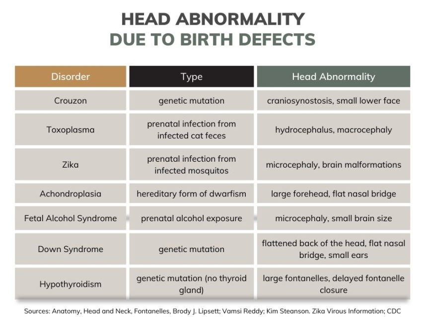
Birth Defects
There are a variety of medical conditions present at birth that are associated with an abnormal head shape. In these cases, the baby’s skull bones do not form properly while growing in utero. This may also be poor formation of nerves and their connections within the brain.
Congenital head abnormalities can occur as a result of genetic mutations, hereditary predisposition, prenatal toxic exposures, and infections. Many are associated with microcephaly or macrocephaly. Abnormalities elsewhere in the body may also be present.
Although there are a vast number of reasons why an infant has a birth defect, a few specific ones are worth mentioning:
What Head Shape Abnormalities Require Treatment?
Most childbirth-related head shape abnormalities self-resolve and do not require treatment. One exception is if a calcified cephalohematoma develops, which causes a significant head deformity. Parents may opt for surgical correction in such cases.
The recommendations for the management of plagiocephaly vary.
In most cases, conservative measures such as more “belly time” and upright positioning as core muscle strength improve are sufficient to promote the return of a normal head shape.
Molding helmets have become a popular “re-shaping” option among parents. Although their use has been endorsed by the American Association of Neurological Surgeons, some clinical studies have not found significant improvements in an infant’s head shape.
If parents choose this type of intervention, the optimal time to seek an evaluation infant is around six months. Waiting until this age is ideal because the head flattening may have already improved or self-resolved. After 18 months old, treatment with a molding helmet can be more difficult and take longer. When an infant’s plagiocephaly is associated with torticolis, physical therapy is the mainstay of treatment. Parents can continue techniques used in therapy sessions at home. Persistent cases require neurology or surgical evaluations.
Head shape abnormalities caused by underlying medical problems often require neurosurgical intervention.
To reduce the increased intracranial pressure of hydrocephalus, a shunt is surgically implanted from the skull to the abdomen in order to drain some of the fluid around the brain.
A brain abscess or collection of blood may require surgical drainage.
Fontanelles that close too soon must be surgically re-opened to allow the infant’s brain to grow normally. Molding helmets may be used after surgery to improve head shape further.
Unfortunately, there is no treatment for microcephaly or the sequelae of many birth defects. Symptoms are managed by a multidisciplinary team that includes neurologists, developmental pediatricians, geneticists, and craniofacial specialists.
Frequently Asked Questions about Baby’s Head Shape
How long does a normal infant’s head take to “round out”?
If areas of a baby’s head have become flattened during the first few months of life, they should naturally begin to “round out” between six to eight months old.
Do babies outgrow torticollis?
Torticollis is not a head shape concern but a head position concern. It is a condition where your baby’s neck muscles cause their head to twist and tilt to one side. So, do babies outgrow torticollis? Yes. Physical therapy is typically initiated by the age of two months, and 97 percent of cases completely resolve. If there is no improvement after six months of physical therapy, botox injections or surgical procedures may be considered.
Is torticollis linked to autism?
No. There is no association with autism.
Do pillows help reduce plagiocephaly?
Whether or not a pillow reduces plagiocephaly has not been studied due to risks to the infant. Pillows, blankets, and other soft surfaces are not recommended for infants and increase the likelihood of sudden infant death syndrome (SIDS).
Will my two-year-old’s head “round out?” At what age is a child’s head shape permanent?
A toddler’s head will continue to reshape itself, albeit at a slower rate, until all of the fontanelles close at age three. The skull bones are also more calcified and have a thicker density at this age.
How common is it for an infant to have a “cone head?”
There are no statistics on the prevalence of temporary molding that occurs during delivery. A caput succedaneum (i.e., cone head”) is seen in up to 33 percent of infants.
Do pediatricians check head shape?
Yes. A baby’s head is examined for any shape abnormalities at each well-visit. Head measurements are recorded on a head circumference growth chart.
What is megalencephaly?
Megalencephaly is the term for an abnormally large brain size in an infant. This differs from macrocephaly which reflects the size of the skull.
Megalencephaly is often associated with malformations of the brain tissue itself or the blood vessels that supply it. As a result, the infant or child may have seizures, spinal cord problems, and speech and motor skills delays. This condition is often a component of a genetic disorder that also affects limbs and other organs.
Are ridges on a baby’s head normal?
Some infants and toddlers have prominent bone edges around the fontanelles that are visible on the skin’s surface. In most cases, these are normal. However, if these ridges are associated with a misshapen head, it is likely that the fontanelles are closing too soon.
Parents should have this evaluated by their pediatrician, and the baby should be referred to a neurosurgeon for imaging.
Takeaway
As seen, there are many birth-related, position-related, and medically-related reasons why a baby’s head shape may be uneven. It is important to remember that in most cases, the head shape abnormalities self-resolve and do not require treatment. However, since not all abnormalities self-resolve and some cases of uneven head shapes are due to medical conditions that need treatment and attention, it is important to consult a pediatrician if you notice anything unusual about your baby’s head shape.
Read Next
- Did I Hurt Baby’s Fontanelle By Accident? 4 Warning Signs and Fontanelle Facts
- Sunken Fontanel by Pulling Out Nipple: The Mollera Caida Myth
Research References
- Anatomy, Head and Neck, Fontanelles – NCBI Bookshelf
- Birth Defects | Zika virus | CDC
- Epidemiology of Positional Plagiocephaly in Children – The ISPN Guide to Pediatric Neurosurgery
- Caput Succedaneum – NCBI Bookshelf
- Macrocephaly – NCBI Bookshelf
- Congenital Torticollis – NCBI Bookshelf

Paula Dennholt founded Easy Baby Life in 2006 and has been a passionate parenting and pregnancy writer since then. Her parenting approach and writing are based on studies in cognitive-behavioral models and therapy for children and her experience as a mother and stepmother. Life as a parent has convinced her of how crucial it is to put relationships before rules. She strongly believes in positive parenting and a science-based approach.
Paula cooperates with a team of pediatricians who assist in reviewing and writing articles.